Plant substances as potential medicines
Purpose-
The purpose of this lab was to find out what plant materials found locally contain active ingredients that will inhibit the growth of bacteria.
Materials-
Procedure-
part1: preparing agar plates
1.Prepare a nutrient or LB broth for the E. coli at least 24 hours in advance. Using sterile technique, add colony of E. coli culture to broth, incubate, then shake at 37*C for 24 hours.
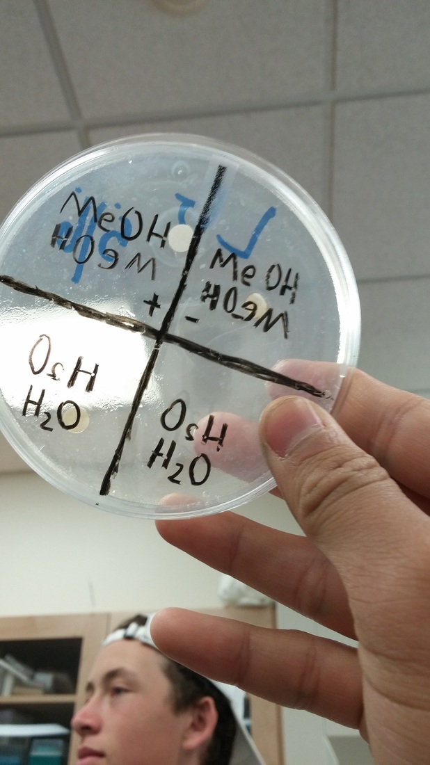
2.Draw +sign on bottom of plate and divide plate into 4 quadrants. Label plate with identification.
3.Liquefy sterile LB agar in the microwave at 50% power. Using sterile technique,pour 20mL of LB into the plate. Let it solidify for 15min. Let dry for at least 24hours.
part2:
4. Grind 2 g of plant tissue (leaves) with 10 mL of deionized water in mortar and pestle, and let sit for 3 minutes. Channel in 11 cm pipe, sanitize extricate with syringe channel, and gather 1 mL of concentrate in marked 1.7 mL microtube.
5. Rehash Step 4, with the exception of supplant deionized water with methanol. Place 1.7 mL tube with 1 mL of methanol concentrate in 65*C warmth piece (tops open) for 24 hours to vanish methanol. Reconstitute dry matter in microtube with 1 mL of deionized water.
6. Rehash Step 4 and 5 for six examples and mark them.
7. Drop channel paper circles in every sifted concentrate tube utilizing sterile forceps (disinfected by being flared in liquor).
8. Set up three negative control circles of just methanol and sterile and refined water.
9. Set up six positive control plates of ampicillin arrangement.
10. Permit plates to be soaked with the concentrate (maybe overnight).
11. Close tubes. Store all specimens at 4*C until prepared to utilize.
part3:
12. Use a clean pipet to move 1 ml of E.coli broth to the middle of a petri dish. sterilize a spreading loop with fire and alcohol, to spread the bacteria evenly. Cover, and leave for at least 15 minutes.
13. Using forceps, place 1 dish in each quadrant, 2 cm from the edge of the petri dish. Place the methanol samples in one dish and the water samples on another.
14. Repeat step 13, in order to end up with 3 methanol and 3 water replicates.
15. Place a negative control disk in the center of the appropriate plate. Then a positive control with amplicillin in a quadrant on each plate.
16. In the end you should have 6 petri dishes with a negative control in the middle and a a positve control and three sample disks.
17. Guarantee that the disk hold fast to surface of agar. Alter the plates and brood at 37*C for 24 to 48 hours.
18. After incubation, search for at the plates with plant concentrate circles for zones of inhibition, clear rasnge shaped by inhibitory (abatement in real life) activity of a substance in the plant material around the circle. Photo the plates, marking any restraint of bacterial development.
19. Make an information table for the repeats and midpoints. Incorporate depictions of the bacterial garden around every circle. Record the breadth and clarity of any cleared zones around the circles in quantitative estimations.
Reflection-
The purpose of this lab was to find out what plant materials found locally contain active ingredients that will inhibit the growth of bacteria.
Materials-
- balance
- LB broth base
- media bottles, 250mL
- sterilizer
- water bath, shaking
- sterile LB agar
- Laminar flow hood and disinfectant
- safety glasses
- bunsen burner
- inoculating loop, Ni/Cr wire
- Petri dishes, sterile
- E. coli
- Plant specimen
- Mortar and pestle
- Pipet, 10mL and pump
- plastic funnel
- filter paper disks
- beakers, 100mL
- syringe and filter
- reaction tubes and rack
- methanol, absolute
- pipet, 1mL and pump
- dry blockheater/heat block
- forceps
- Ampicillin
- glass spreader
- incubator oven
Procedure-
part1: preparing agar plates
1.Prepare a nutrient or LB broth for the E. coli at least 24 hours in advance. Using sterile technique, add colony of E. coli culture to broth, incubate, then shake at 37*C for 24 hours.
2.Draw +sign on bottom of plate and divide plate into 4 quadrants. Label plate with identification.
3.Liquefy sterile LB agar in the microwave at 50% power. Using sterile technique,pour 20mL of LB into the plate. Let it solidify for 15min. Let dry for at least 24hours.
part2:
4. Grind 2 g of plant tissue (leaves) with 10 mL of deionized water in mortar and pestle, and let sit for 3 minutes. Channel in 11 cm pipe, sanitize extricate with syringe channel, and gather 1 mL of concentrate in marked 1.7 mL microtube.
5. Rehash Step 4, with the exception of supplant deionized water with methanol. Place 1.7 mL tube with 1 mL of methanol concentrate in 65*C warmth piece (tops open) for 24 hours to vanish methanol. Reconstitute dry matter in microtube with 1 mL of deionized water.
6. Rehash Step 4 and 5 for six examples and mark them.
7. Drop channel paper circles in every sifted concentrate tube utilizing sterile forceps (disinfected by being flared in liquor).
8. Set up three negative control circles of just methanol and sterile and refined water.
9. Set up six positive control plates of ampicillin arrangement.
10. Permit plates to be soaked with the concentrate (maybe overnight).
11. Close tubes. Store all specimens at 4*C until prepared to utilize.
part3:
12. Use a clean pipet to move 1 ml of E.coli broth to the middle of a petri dish. sterilize a spreading loop with fire and alcohol, to spread the bacteria evenly. Cover, and leave for at least 15 minutes.
13. Using forceps, place 1 dish in each quadrant, 2 cm from the edge of the petri dish. Place the methanol samples in one dish and the water samples on another.
14. Repeat step 13, in order to end up with 3 methanol and 3 water replicates.
15. Place a negative control disk in the center of the appropriate plate. Then a positive control with amplicillin in a quadrant on each plate.
16. In the end you should have 6 petri dishes with a negative control in the middle and a a positve control and three sample disks.
17. Guarantee that the disk hold fast to surface of agar. Alter the plates and brood at 37*C for 24 to 48 hours.
18. After incubation, search for at the plates with plant concentrate circles for zones of inhibition, clear rasnge shaped by inhibitory (abatement in real life) activity of a substance in the plant material around the circle. Photo the plates, marking any restraint of bacterial development.
19. Make an information table for the repeats and midpoints. Incorporate depictions of the bacterial garden around every circle. Record the breadth and clarity of any cleared zones around the circles in quantitative estimations.
Reflection-